Mri Pelvic Floor Radiographics

Pelvic Floor Failure Mr Imaging Evaluation Of Anatomic And Functional Abnormalities Radiographics

Great Pelvic Compartment Diagram By Dr Vikas Shah Radiologist And Apops Medical Advisory Committee Member Pelvic Floor Abdominal Pelvic Organ Prolapse

Mri Defaecating Proctogram The Xray Doctor Mri Health Articles Mens Health
Https Pubs Rsna Org Doi Pdf 10 1148 Rg 345140137

Mri Of The Male Pelvic Floor Radiographics

Pin By Dr Vikas Shah On Thexraydoctor Rectal Prolapse Rectal Pelvic Floor

Mr imaging based assessment of the female pelvic floor.
Mri pelvic floor radiographics.

Magnetic Resonance Imaging Of The Pelvic Floor Sciencedirect
Mri Pelvic Floor

Pin On Musculoskeletal Radiology

Dynamic Mri Of The Pelvic Floor Open Mri Manchester

Normal And Variant Pelvic Anatomy On Mri Radiology Key

Abdominal Xray Showing Thumbprinting Of Large Bowel A Sign Of Colitis Radiology Imaging Foamrad Foamed Large Bowel Radiology Figure Painting

Bilateral Hydronephrosis And Megaureters Due To Grade Iii Cystocele Amp Grade Iv Total Uterine Prolapse

Sagittal T2 Weighted Image Of An Mri Of The Lumbar Spine Lumbar Spine Mri Anatomy This Is A Commonly Perf Radiology Student Medical Anatomy Anatomy

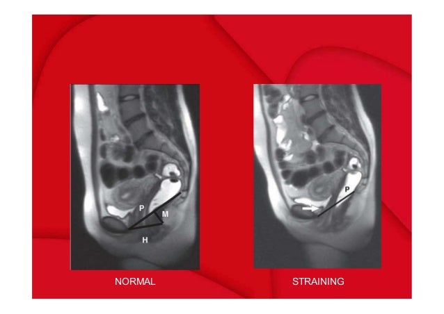
Mri For Pelvic Floor Dysfunction Can The Strain Phase Be Eliminated Semantic Scholar

Abdomen Anatomy Mri Abdomen Axial Anatomy Free Cross Sectional Anatomy Abdominal Aorta Superior Mesenteric Artery Rectus Abdominis Muscle

Abdomen X Ray

Adenoid And Palatine Tonsil On Lateral Radiograph Radiology Case Radiopaedia Org Radiographer Radiology Medical Knowledge

Bringing Some Hollywood And Oscar Night Glitz And Glamour To Radiology False Toenails Fashion Instagram Posts Instagram
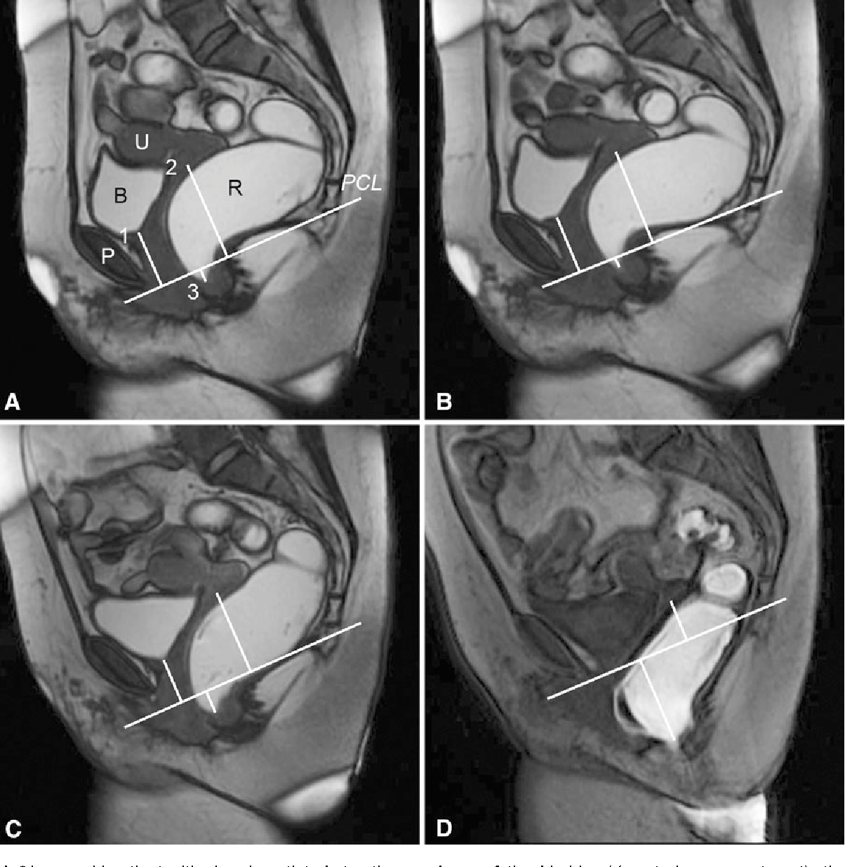
Figure 3 From Dynamic Pelvic Floor Imaging Mri Techniques And Imaging Parameters Semantic Scholar

Abdominal X Ray

Pin On Thexraydoctor

Ischium Wikipedia Pelvic Girdle Pelvic Floor Muscles Pelvic Floor

Pin On Brain

Pin By Saeed Esmailian On Cns Radiology Imaging Radiology Pet Ct

Magnetic Resonance Imaging Of The Female Pelvic Floor Radiology Key
Pelvic Floor Lipoma Radiology Case Radiopaedia Org
Take A Look At This Annotated X Ray Which Exhibits You The Place A Number Of The Pelvic Musc Medical Anatomy Radiology Student Radiology

Normal Radiographic Anatomy Of The Knee Radiology Case

Dynamic Magnetic Resonance Imaging For Assessment Of Minimally Invasive Pelvic Floor Reconstruction With Polypropylene Implant European Journal Of Radiology
Source : pinterest.com
